For many patients, corneal ectasia does not begin with a dramatic event. It begins with vision that seems to keep changing, glasses that no longer feel reliable, more glare at night, or increasing irregular astigmatism. In some cases, ectasia is related to an underlying corneal condition. In others, it may develop after refractive surgery such as LASIK or PRK.
This page is designed as a starting point. It explains the condition in clear language, outlines how specialists diagnose it, and helps patients understand the broad treatment paths available today before moving into more detailed treatment-specific pages.
What is Corneal Ectasia, and why it affects vision.
Common symptoms and how they often begin.
Possible causes, risks, factors, behaviours.
How doctors confirm the diagnosys.
What treatment options are available?
What results can you expect after treatment.
Visão geral
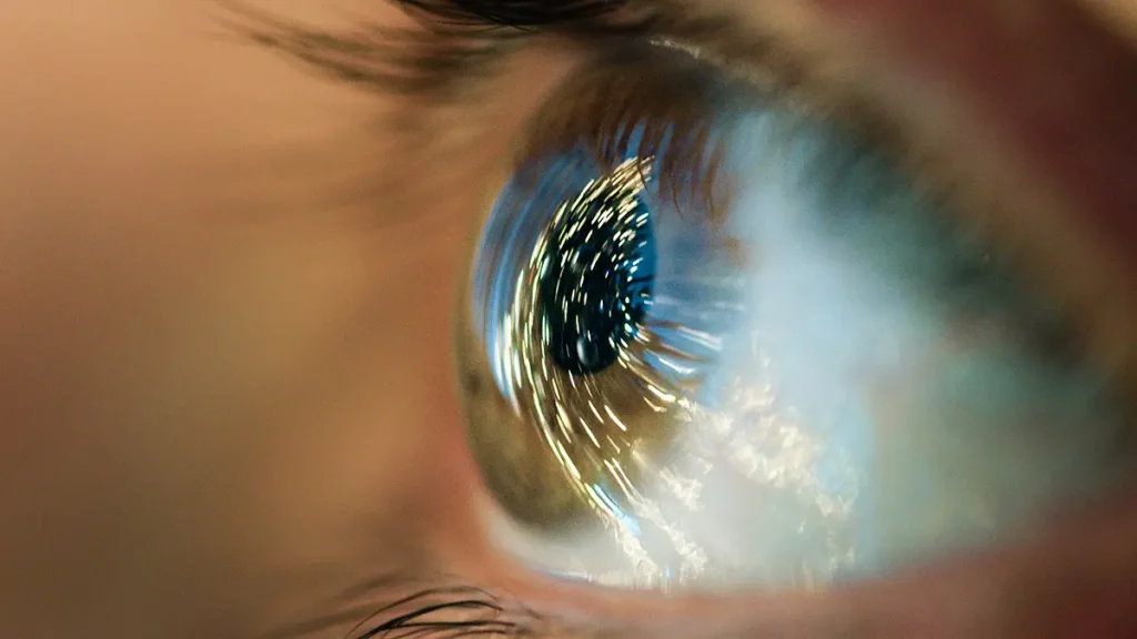
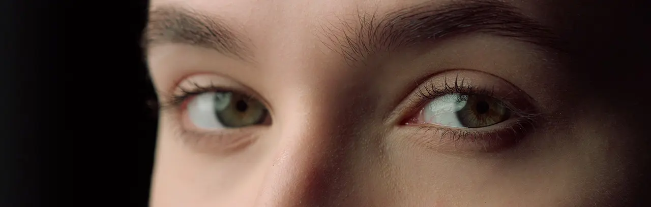
Corneal ectasia is not one single disease. It is an umbrella term for a group of disorders in which the cornea becomes thinner, weaker, and more irregular in shape. As the cornea bulges outward, it can create increasing astigmatism and reduce the sharpness and quality of vision over time. The degree of visual impairment can vary widely, from mild changes to significant loss of corrected visual quality.
This group includes conditions such as keratoconus, pellucid marginal degeneration, and keratoglobus, as well as ectasia that can develop after refractive surgery. Because these conditions share structural weakening of the cornea, they are often assessed and managed within the same broader corneal framework.
Sintomas
Symptoms often develop gradually, which is one reason patients may spend time changing prescriptions before receiving a precise diagnosis.
The most common symptom is progressive visual decline over time, even when glasses or contact lenses are updated.
Many patients notice more glare, halos around lights, and greater difficulty seeing at night.
As the cornea becomes more irregular, astigmatism may become harder to correct with standard glasses.
The most common symptom is progressive visual decline over time, even when glasses or contact lenses are updated.
Causas
The cause depends on the type of ectasia. Some forms happen naturally because the cornea is structurally predisposed to thinning and bulging. Others develop after surgery because the cornea was already vulnerable or because its biomechanical strength was reduced beyond what it could safely tolerate.
In naturally occurring ectatic disease, family history can matter. In both keratoconus-related and other ectatic patterns, chronic eye rubbing and conditions associated with itching or irritation may also contribute to risk or progression. In post-refractive ectasia, careful screening with corneal imaging is important because subtle pre-existing risk factors may not always be obvious without detailed testing.
If you have corneal ectasia or are being evaluated for it, avoiding eye rubbing is commonly advised.
A form of corneal ectasia can develop after refractive procedures such as LASIK, and it has also been reported after PRK and other corneal refractive surgeries. It may appear early, but it can also be diagnosed months or even years later. Patients may notice increasing myopia, increasing astigmatism, worsening uncorrected vision, or a loss of best-corrected vision after initially good surgical results.
This does not mean refractive surgery is broadly unsafe. It means that corneal screening matters. Modern preoperative assessment uses topography, tomography, pachymetry, and related data to identify corneas that may be at higher risk before surgery is considered.
Diagnosis
Diagnosis usually begins with a comprehensive eye examination, a discussion of symptoms and history, and specialized corneal imaging. Specialists look not only at how well you see, but also at the shape, thickness, and structural behavior of the cornea. Corneal topography and tomography are central because they help reveal irregular steepening, asymmetry, posterior elevation, and other patterns that are important in ectatic disease.
This helps show how much the corneal changes are affecting visual quality.
A specialist evaluates the front of the eye and looks for signs associated with ectatic disease.
Topography and tomography show the shape of the cornea in detail and can reveal patterns consistent with ectasia.
Pachymetry and repeat scans over time help determine whether the cornea is stable or changing. This is especially important when treatment decisions depend on progression.
A good diagnosis does more than name the condition. It helps define the type of ectasia, its severity, whether it is progressing, and which treatment path may be most appropriate.
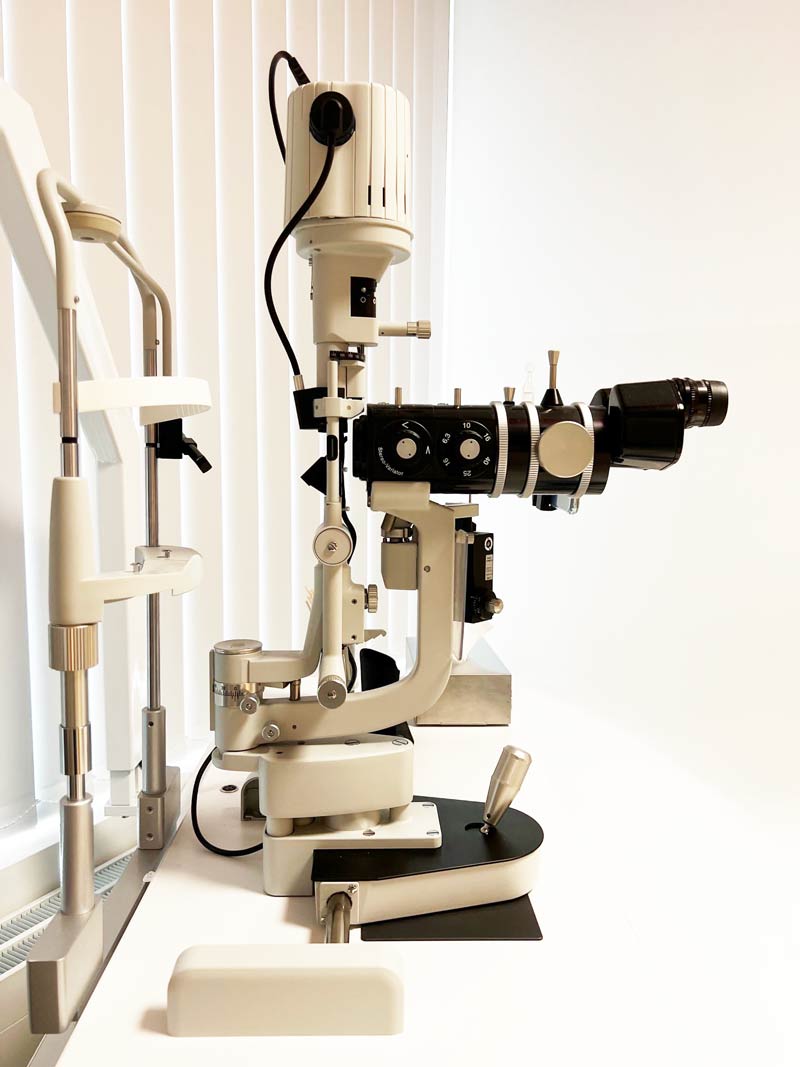
Observamos de perto a sua córnea através do biomicroscópio e avaliamos a sua prescrição e se o seu astigmatismo se alterou ao longo do tempo.
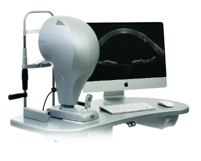
Um exame essencial que pode detetar mesmo as fases iniciais da doença. É criado um mapa detalhado da córnea, do seu epitélio e da sua espessura. Utilizamos a variante mais moderna, combinando a tomografia de coerência ótica (OCT) e medições baseadas em placido (MS-39).
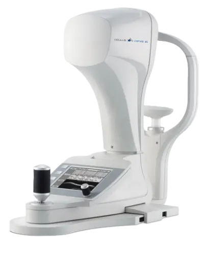
Utilizamos um sofisticado sistema de imagem Scheimpflug que capta mais de 4.000 imagens por segundo para analisar a forma como a sua córnea reage biomecanicamente (CosVis ST).
Treatment
Treatment depends on the type of ectasia, how advanced it is, whether it is progressing, and how much vision is affected. In general, treatment falls into two broad goals: improving visual quality and helping stabilize the cornea.
Outcomes
Outcomes vary. Much depends on the type of ectasia, how early it is diagnosed, whether it is progressing, and which treatment path is chosen. Many patients can maintain or regain useful functional vision with the right combination of monitoring, vision correction, and corneal treatment. Appropriate management can often reduce the need for transplantation, which is generally reserved for more advanced cases.
For patients, success is not only about scans or measurements. It is also about practical life: clearer vision, more confidence with driving and screens, a better understanding of what is happening, and a treatment plan that makes sense for the stage of the condition. This is an inference based on the goals of ectasia management, which centers on visual function and stabilization.
Porquê nós?
Corneal ectasia requires more than a routine eye exam. It requires precise diagnostics, experience with complex corneas, and a team that understands both naturally occurring ectatic disease and ectasia after LASIK, PRK, or SMILE. The right treatment depends on understanding exactly why the cornea has changed, whether the condition is still progressing, and which option offers the best chance of stabilizing vision
ELZA has pioneering role in post-refractive ectasia and corneal cross-linking, has internationally recognized corneal specialists, and offers strong support for international patients.
For patients, that means careful evaluation, realistic guidance, and treatment planning built around the best achievable visual outcome.
For many patients, success means reading more comfortably, using screens more easily, and feeling more confident in daily life.
The right treatment begins with precise analysis of corneal shape, thickness, and progression risk. This is critical in both natural ectatic disease and ectasia after surgery.
ELZA states that its team includes internationally recognized leaders in ophthalmology and corneal cross-linking.
The focus is on stabilizing the cornea, preserving or improving functional vision, and guiding each patient toward the most appropriate next step.
Online consultations and dedicated international-patient pathways make specialist input easier to access from abroad.
FAQs
Not exactly. Keratoconus is one type of corneal ectasia, but the term “corneal ectasia” is broader and also includes other thinning-and-bulging disorders as well as ectasia that can happen after refractive surgery.
Yes. Post-refractive ectasia can appear months to years after surgery, which is why long-term changes in vision after LASIK or PRK should be evaluated properly.
They help improve vision, but they do not strengthen the cornea or stop progression on their own. That is why progression assessment is so important.
No. Some patients are monitored, and some do well with glasses or specialty contact lenses. Surgical treatment is considered when progression is documented or when visual quality cannot be adequately managed more conservatively.
Yes. Cross-linking is used to strengthen corneal tissue and is recommended for progressive ectatic disease because its main goal is to stabilize the cornea.
Entrar em contacto
Durante o horário de expediente.
Envie-nos um e-mail.
Marque uma consulta e venha ver-nos.
Obrigado por escrever uma avaliação no Google.
Contacte-nos aqui e entraremos em contacto consigo.
Consulta online com zoom para os nossos pacientes internacionais.
Contacte-nos aqui e entraremos em contacto consigo.
Contacte-nos aqui e entraremos em contacto consigo.
Mantenha-se informado & receba o boletim informativo
Aderiu com sucesso à nossa lista de subscritores.
Receber e informar sobre o boletim informativo
Eles estão a ser bem sucedidos na nossa lista de contactos.
Por favor, melhore a sua assinatura, ao clicar no link do e-mail, que nós já enviámos.